Nomenclature
Speaking of helixes, and browsing through the bibliography, it is quite clear that the terms used to name the two kinds of helices, inverted in a mirror, can be confusing: right and left, as well as clock-wise or anti-clockwise.
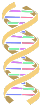
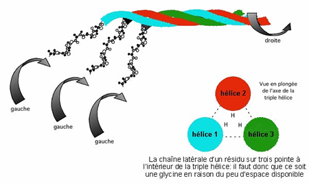
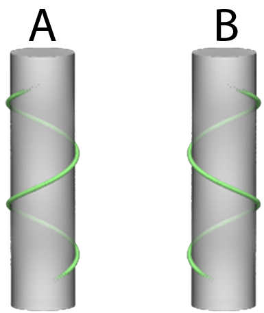
In the illustration bellow, a DNA helix named Right, then a collagen molecule, also named Right helix and composed of 3 strands of pro-collagen, which are formed in a helix named left. This is the official nomenclature against which we will take issue for obvious reasons of understanding how the patient’s body works. |
|
|
« Official » nomenclature A : Right helix ; B : Left helix |
|
This official nomenclature refers to the observer. If we imagine that the helix named A is a screw, I will have to turn my screwdriver towards the right to screw it and it will be the opposite for the B helix. It is therefore the rotation of the observer’s hand that we are talking about when we say that A is right and B is left.
We have consciously abandoned this official nomenclature for two reasons, one ethical, the other pragmatic.
The first one is that I find it highly inappropriate to name something in the point of view of the observer, it places the zero of space in the observer’s naval and thus testify to an egocentrism, which in my opinion is rather revealing of the society we live in. It seems to me that when we give attention to an object, it should become, the Zero, the centre of our concerns and, as our helix, ultimately will be a patient. It is quite natural to see things from their perspective if we are taking care of them. To define the direction of rotation of a helix, we position ourselves in the axis of the helix, in the patient’s axis. Same thing if it’s not specified when we speak about left and right, it’s the patient’s left and right we are talking about and not the practitioners. It’s the only way to homogenise the observations on bipeds and quadrupeds and not get confused unnecessarily.
The second one is that since this definition has been said, the student’s understanding in the “Tensegrity Body” courses has become much more fluid and there isn’t any confusion about “right/left” to visualise the phenomenon.
Also, for these reasons, we decide to choose our own convention to make things easier for us. We then call the A helix a LEFT helix (L). If we are looking from the patient’s perspective, in the helix’s axal, we can observe that the first bottom spire crosses in front of the helix (ventrally of the patient) from right to left going up (cranially). The B helix becomes RIGHT (R). By specifying L for levogyre, we introduce the notion of movement: life is movement, movement leans on a structure. This term Levogyre, already mimics the physiological torsion [1] which we have already talked about. The hipbone, following the helix movement, goes up on a portion of spire then goes back down and releases itself.
|
|
Osteopathic nomenclature in this concept |
This nomenclature simplifies the understanding for the osteopath. The patient remains the spatial reference point and the direction of the helix is determined by the first coil from the bottom of the patient. Our LEFT helix (L) sees its first turn starting from the bottom, on the ventral side of the patient, and goes from their right side towards their left side and the other way around on the dorsal side. That way, whatever the position of the osteopath in relation to his patient, wherever it’s a biped or quadruped, the helix we are talking about is the same and vocabulary mistakes cannot be done. It eases things for our treatment that we aren’t following the official nomenclature, which is inadequate for understanding the concept and the care that results from it.
Therefore, when you read:
The gastropods shell is said dextrogyre, for the majority of them. If you would put yourself in the snail’s perspective in its shell, you would notice that the first spire from the foot of the snail goes toward the left, direction of the spiral of the snail’s body from the inside. |

|
Vines roll up towards the right (generally speaking) … true for the observer’s point of view, but if the observer puts oneself into the plant’s axal, it is from bottom to top towards the left that they go! |

|
Why a LEFT (L) helix ?
This choice to build a helix with the body and even more, a Left (L) helix is a sensible idea coming from observing our everyday life (a new-born that we rock, etc) but also from a very rationalised matter.
The mathematician Mandelbrot, described a class of mathematical objects, called the fractals [2]. These fractals have something peculiar; they are the repetition of the same figure on several levels. They are said invariable in scale. It has been shown that living organisms rely on such a system to achieve economies of structure and energy, it is the same structure that is repeated on all levels of living organisms. The Romanesco cabbage is multiple cones within a cone.

The sunflower above makes a similar spiral of elements, branching of trees or the lungs arteries tree shaped have been proved to have fractals properties. The bee on the sunflower is composed of metamers, similar particles that then specialise according to where they are in the body.
So, assuming that most of our fascia proteins (collagen in particular) are LEFT helices (L) and that fascia, the most important tissue in our body in terms of structuring the body, is a major concern in osteopathy. We are going to apply the fractal principal and state that to each level of superior organisation we are keeping the LEFT (L) helix structure. The molecules of structures are LEFT (L) helices, cells are organised in LEFT (L) helices, tissues are LEFT (L) helices, organs are LEFT (L) helices, the body is a LEFT (L) helix. It’s not logical at first and it could be an incorrect keyboard short cut. Although:
-
We find a helicoidal structure in the heart wall. A spiral easy to observe at a butcher’s shop when unrolling the bovine’s cardiac muscle.
-
All obstetricians know that to help give birth to a mammal (new-born, foal, calf, etc) the easiest movement makes a helix. It was in Quebec that I first heard the term "birth helix" used to describe this movement.
In doing so, we then adapt the concept of physiological torsion and apply it along an external fascia rail: the fascial helix that wraps the body on its surface and that rests on this fascia. This fascial helix is considered as a rail on which this twisting movement is applied.
This raises two questions :
-
The first one is: can this helix be seen in histology? Can it materialize and be seen directly? For me, the answer is no. Gimbertaud in his films on fascia, shows us how restructuration of fascial tissue is constantly happening and is fundamental in comprehension of its remodelling and movement. So, if there is a helix shape, in my opinion, we must consider it as a dynamic process, next though it needs to be understood that we are not looking for a helix but the capacity for tissues to form a helix. Another way of saying that we are looking for the ability for the body to change in a helical manner.
-
The second one is: Is there a RIGHT helix? Again, for me, the answer is no, not like the equivalent of a LEFT (L) helix. Putting back a helix straight would restore the symmetry we all know, but should exist, for me, only to satisfy us into the comfort of our thought. We need to think asymmetrically to be efficient. Let’s not forget that the molecule of collagen, which is a LEFT (L) helix, is the sum of three procollagen molecules which are RIGHT (R) helices. Therefore, the RIGHT (R) helix is a constituent of the LEFT (L) helix of collagen. DNA, which is a LEFT (L) helix under the A or B form which are the most common, is a RIGHT (R) helix under the Z shape when it is undergoing transcriptional activity. Cross helicoidal patterns, alternating right and left helices to different levels, are found invariably in scale in living structures. “Such crossed helical patterns in the muscles and fascia surrounding the spine and synovial joints are likely to contribute to their flexibility and stability" [3].
Conclusion
The helicoidal structure appears scale-invariant from macrocosm to microcosm. The majority of observed helices seem to be LEFT helices (L): the majority of described galaxies are spiralized in LEFT helix (L), numerous plants, our body (global movement facilitated following the rotation of a LEFT helix (L), some organs like the heart, the majority of helicoidal molecules of structure (collagen), DNA, … on the other note, these LEFT helices (L) can be made out of right helices which helps contributes to certain properties of deformability for example. Here again the L/R dissymmetry appears. To clarify the nomenclature this way makes it possible to find our way around the various tests and osteopathic treatments proposed to treat the dysfunctions generated by this system.