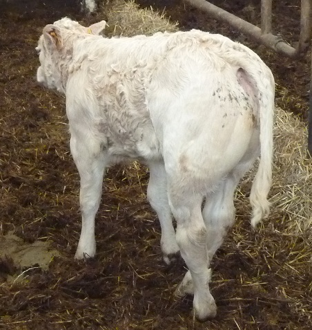
A two month old calf got sent to me by his veterinarian for a hind leg (right one) lameness, or hind legs ataxia. When he was 15 days old, this calf had an ombilical infection and receved antibiotics and anti-inflammatories. The lameness occured one week after the start of the navel infection and progressively got worse through time. Despite the healing of the navel, the lameness continues while anti-inflammatories and antibiotics are given over 6 weeks, without any effects, even with change of molecules classes.
Consultation
On examination, the calf shows hesitating walking, quite ataxic and with hindlegs crossing. The right hindleg is always put on the ground medialy, under the abdomen. The sacrum is moved towards the right, pulling the start of the tail, which only wags to the right.
On palpatory examination, the calf shows an important hot area in L3-L4.
On osteopathic examination, the calf shows three dysfuntions chains, all of them starting at the ombilic.
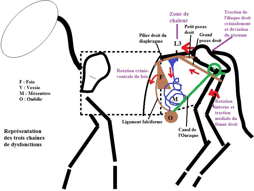
The first one:
– it deals in the first place with the falciform ligament, which excessive tension pulls the liver into a cranio-ventral rotation.
– the liver then, pulls on its attachments : the coronary ligaments on one side, which hold it on the spine while joining the diaphragm poles, and the triangular ligaments on the other side, directly linked to the diaphragm.
– Unbalance of the diaphgram creates a strong traction on the right diaphragm pole, which slides between the psoas opposite L3, and puts them into dysfunction as well using mechanical disturbance. Knowing that the big psoas goes from under the transversal lumbar processus to the cranio-medial face of the femur, its contracture explains the fact that the right hindleg is hold medialy.
The small psoas goes from the vertebral bodies of the first lumbar vertebras and finishes cranialy on the coxal bone. Its contracture creates a cranial traction of the ilium, which pulls the sacrum, explaining the visible right deviation of the sacrum apex and the start of the tail.
It is then through a tensegrity game of abnormal forces lines, starting at the ombilic and reaching the right hind leg, that the calf is lame. And its lameness is really directly linkable to its infected navel, the infection having left behind healing tensions, which, through the liver, enhances the right paralumbar muscles traction.
The second chain of dysfunction :
– still starts at the ombilic, but this time goes through the peritoneum and the mesentere. The ombilical infection has probably started a localised peritonitis, with parts of mesentere sticking to the abdominal wall : the mesentere being attached to the dorsal part of the abdominal wall facing the cranial under-lumbar area.
This area, then, recieves two lines of abnormal forces, which can explain the probable L3-L4 disc compression, which can be felt through tissular technics, and through palpation of the strong heat of this area. This discal compression and the associated tissular tensions around the nerves roots, explain the calf hind leg ataxia.
Finaly a third dysfunctionnal chain :
Of ombilical origin, this time, it goes caudaly, throuh the Urachus canal, and induces a medial rotation of the bladder. This last chain of tensions ends up on the pubis, and doing that, finishes unbalancing the coxal bones.
After the osteopathic consultation,
The calf ataxia regresses progressively. The improvement is really noticeable 8 days later, and at ten days, the calf is jumping in his stable without showing any sign of his ancient lameness. The tail is back to its medial position and moves freely on both sides. The right hind leg has found its normal position again.
Conclusion.
This clinical case illustrates in bovine practice the complementary action of osteopathy and allopathy : when the allopath has a chemical vision of the diseases, the osteopath gives a reading of the physical forces in the body, which can bring a different understanding of certain diseases and even their occurences.
Here, the ombilical infection could not have been treated without antibiotics, but allopathy could not do everything for the lameness. The osteopath could not have treated the infection, but gave the opportunity to understand and treat the lameness built up by healing tensions.
This calf has been cured thanks to the synergetic use of both medecines.